38 picture of the eye with labels
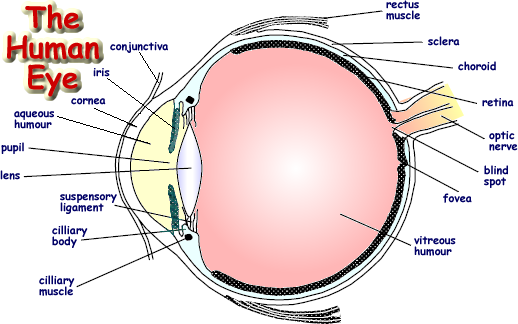
Eye Diagram With Labels and detailed description - BYJUS Iris is the coloured part of the eye and controls the amount of light entering the eye by regulating the size of the pupil. The lens is located just behind the iris. Its function is to focus the light on the retina. The optic nerve transmits electrical signals from the retina to the brain. Pupil is the opening at the centre of the iris. Label Eye Printout - EnchantedLearning.com Label the Eye Diagram. Human Anatomy. Read the definitions, then label the eye anatomy diagram below. Cornea - the clear, dome-shaped tissue covering the front of the eye. Iris - the colored part of the eye - it controls the amount of light that enters the eye by changing the size of the pupil. Lens - a crystalline structure located just behind ...
Eye Anatomy: A Closer Look At the Parts of the Eye - All About Vision The eye's crystalline lens is located directly behind the pupil and further focuses light. Through a process called accommodation, this lens helps the eye automatically focus on near and approaching objects, like an autofocus camera lens. ... The retina acts like an electronic image sensor of a digital camera, converting optical images into ...
Picture of the eye with labels
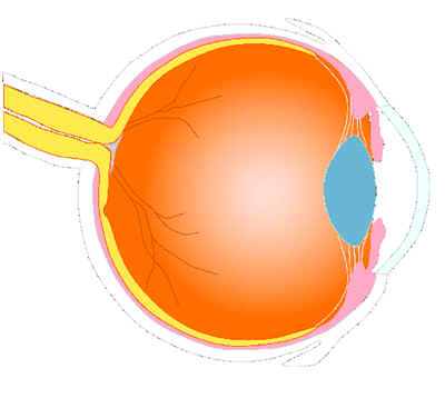
Human Eye Coloring Page | crayola.com The eye is the organ that collects images and sends them to the brain, so you can see. The eye is protected by the bones of your skull and six muscles. Light comes through the pupil which causes the cornea and lens to focus on an image. When the image is projected through the eye, onto the retina wall, the image appears upside down. Eye ... Eye Anatomy Detail Picture Image on MedicineNet.com Cornea: clear front window of the eye that transmits and focuses light into the eye. Iris: colored part of the eye that helps regulate the amount of light that enters. Pupil: dark aperture in the iris that determines how much light is let into the eye. Lens: transparent structure inside the eye that focuses light rays onto the retina. PDF Eye Anatomy Handout - National Institutes of Health of light entering the eye. Lens: The lens is a clear part of the eye behind the iris that helps to focus light, or an image, on the retina. Macula: The macula is the small, sensitive area of the retina that gives central vision. It is located in the center of the retina. Optic nerve: The optic nerve is the largest sensory nerve of the eye.
Picture of the eye with labels. Label Functions of Parts of the Human Eye - University of Dayton Functions of the Parts of the Eye. Select the correct label for the function of each part of the eye. The image is taken from above the left eye. Click on the Score button to see how you did. Incorrect answers will be marked in red. Diagram of the Eye - Lions Eye Institute The eye - one of the most complex organisms in the human body. ... When light hits the retina, a picture travels through the optic nerve to the brain. Optic-Nerve. The optic nerve is a thick bundle of nerve fibers that connect the back of the eye (retina) to the brain. It transfers all the visual information to the brain which then interprets ... Label Parts of the Human Eye - University of Dayton Parts of the Eye. Select the correct label for each part of the eye. The image is taken from above the left eye. Click on the Score button to see how you did. Incorrect answers will be marked in red. ... Eye Anatomy: 16 Parts of the Eye & Their Functions - Vision Center The following are parts of the human eyes and their functions: 1. Conjunctiva. The conjunctiva is the membrane covering the sclera (white portion of your eye). The conjunctiva also covers the interior of your eyelids. Conjunctivitis, often known as pink eye, occurs when this thin membrane becomes inflamed or swollen.
60,892 Human eye anatomy Images, Stock Photos & Vectors - Shutterstock Find Human eye anatomy stock images in HD and millions of other royalty-free stock photos, illustrations and vectors in the Shutterstock collection. Thousands of new, high-quality pictures added every day. What is an eye mark and why do I need it? - Consolidated Label An 'eye mark' (also known as 'eye spot') is a small rectangular printed area located near the edge of the printed flexible packaging material. ... Consolidated Label. 2001 E Lake Mary Blvd Sanford, FL 32773. Local: 1-407-339-2626 Toll Free: 1-800-475-2235 Fax: 1-407-331-1711 Label the Eye - The Biology Corner Label the Eye. Shannan Muskopf December 30, 2019. This worksheet shows an image of the eye with structures numbered. Students practice labeling the eye or teachers can print this to use as an assessment. There are two versions on the google doc and pdf file, one where the word bank is included and another with no word bank for differentiation. The Human Eye (Eyeball) Diagram, Parts and Pictures The human eye consists of the eyeball, optic nerve, orbit and appendages (eyelids, extraocular muscles and lacrimal glands). While the eyeball is the actual sensory organ, the other parts of of the eye are equally important in maintaining the health and function of the eye as a whole. The structure of the human eye is such that light can enter ...
Eye Anatomy Diagram - EnchantedLearning.com Aqueous humor - the clear, watery fluid inside the eye. It provides nutrients to the eye. Astigmatism - a condition in which the lens is warped, causing images not to focus properly on the retina. Binocular vision - the coordinated use of two eyes which gives the ability to see the world in three dimensions - 3D. Cones - cells the in the retina that sense color. 31 Most Beautiful Eyes in the World - Woman's World Getty Images. Eyes are naturally beautiful — from the delicate shapes and unique colors to the countless expressions that can be made with them. Blue eyes, brown eyes, green eyes, hazel eyes, gray eyes, and any shade in between are all stunning. Please don't ask us to pick a favorite! Human Ear Diagram Without Labels - picture front of the eye without ... Human Ear Diagram Without Labels - 18 images - circulatory system diagram without labels fresh smorgasbord variety, exam macros trauma yellow, datei anatomy of the human ear svg wikipedia, datei anatomy of the human ear svg wikipedia, Structure and Functions of Human Eye with labelled Diagram - BYJUS The External Structure of an Eye. Sclera: It is a white visible portion. It is made up of dense connective tissue and protects the inner parts. Conjunctiva: It lines the sclera and is made up of stratified squamous epithelium. It keeps our eyes moist and clear and provides lubrication by secreting mucus and tears.
picture front of the eye without labels clipart 20 free Cliparts | Download images on Clipground ...
Labelled Diagram of Human Eye, Explanation and Function - VEDANTU Function of Lens in the Human Eye. The main function of this lens is to focus the light rays that come into our eyes.The lens may be a transparent flexible tissue located directly behind the iris and therefore the pupil. To focus light and images on the retina becomes the basic function of the lens. The cornea and the lens are responsible for ...

Premium Flowers: The cascade wedding bouquet
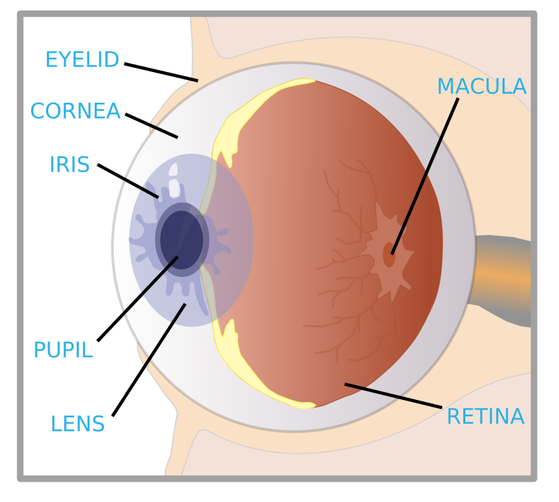
A Picture of the Eye - WebMD The front part (what you see in the mirror) includes: Iris: the colored part. Cornea: a clear dome over the iris. Pupil: the black circular opening in the iris that lets light in. Sclera: the ...

BB CUTE WORLD: Natalie Glebova (Russia)
Human Eye Anatomy Pictures, Images and Stock Photos Browse 7,853 human eye anatomy stock photos and images available, or search for vision or retina to find more great stock photos and pictures. Newest results. vision. retina. human eye structure. eye chart.
Label The Eye - ClipArt Best
What Does the Eye Look Like? - Harvard Eye Associates It is mostly water and gives the eye its form and shape. Our eyes are vital for seeing the world around us. Keep them healthy by maintaining regular vision exams. Contact Harvard Eye Associates at 949-951-2020 or harvardeye.com to schedule an appointment today.

Label The Eye - ClipArt Best
PDF Parts of the Eye - National Institutes of Health Eye Diagram Handout Author: National Eye Health Education Program of the National Eye Institute, National Institutes of Health Subject: Handout illustrating parts of the eye Keywords: parts of the eye, eye diagram, vitreous gel, iris, cornea, pupil, lens, optic nerve, macula, retina Created Date: 12/16/2011 12:39:09 PM

Popular Male Model Yvan Cournoyer | Model Galleries
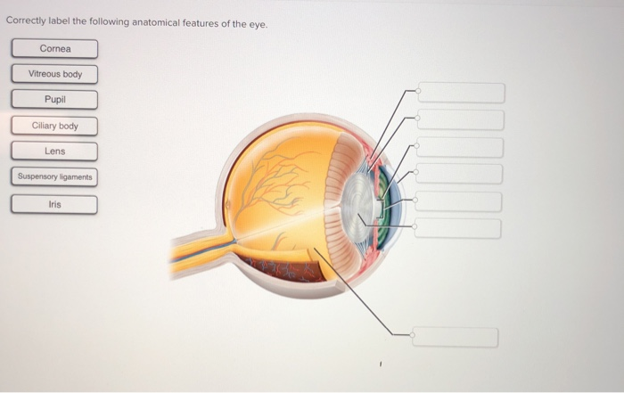
Solved B с A E F D Match the following parts of the eye with | Chegg.com Science. Anatomy and Physiology. Anatomy and Physiology questions and answers. B с A E F D Match the following parts of the eye with the labels in the picture above. A Iris F Cornea В. Ciliary Muscles G Optic Nerve C Lens E Retina Aqueous and Vitreous Fluid. Question: B с A E F D Match the following parts of the eye with the labels in the ...

Label The Eye - YouTube
Transverse section of eye anatomy with labels. - Getty Images View top-quality illustrations of Transverse Section Of Eye Anatomy With Labels. Find premium, high-resolution illustrative art at Getty Images.
33 Label Of The Eye - Labels For You
Human eye diagram, Eye anatomy, Diagram of the eye - Pinterest Science Notes. CONTENTSEyesVideo: Anatomy and Function of the EyeEarsVideo: Ear Anatomy Our most important sensory receptors are the eyes and the ears. The eye is the primary organ for sight, and the ear is the primary organ for sound and equilibrium. Obviously, any impairment of either of these sensory receptors can be a traumatic experience ...

32 Label Human Eye - Labels For Your Ideas
Labelling the eye — Science Learning Hub Labelling the eye. Use this interactive to label different parts of the human eye. Drag and drop the text labels onto the boxes next to the diagram. Selecting or hovering over a box will highlight each area in the diagram. The human eye has several structures that enable entering light energy to be converted to electrochemical energy.

Muscle gallery: muscular black
Quiz: Label The Parts Of The Eye - ProProfs Quiz How much did you get to understand about the human eye? Take up this quiz and find out! Questions and Answers. 1. A is pointing to what part of the eye? A. Cornea. B. Optic Nerve.

New Art Funny Wallpapers Jokes: Beautiful Attractive Eyes of Girls 1440x900 Your Desktop Wallpapers
30 Eye-Catching Wine Label Designs For Inspiration The designer used squares to provide the information. 02. The Cloud Factory. The Cloud Factory wine label design looks simple because of its use of two colors only. Yellow and white colors give the label a soft look, which seems to be the intention of the designer and the brand. 03.
eye with labels by ryanlerch - an image originally sourced from the US government EPA "Sunwise ...
PDF Eye Anatomy Handout - National Institutes of Health of light entering the eye. Lens: The lens is a clear part of the eye behind the iris that helps to focus light, or an image, on the retina. Macula: The macula is the small, sensitive area of the retina that gives central vision. It is located in the center of the retina. Optic nerve: The optic nerve is the largest sensory nerve of the eye.

Eye Label
Eye Anatomy Detail Picture Image on MedicineNet.com Cornea: clear front window of the eye that transmits and focuses light into the eye. Iris: colored part of the eye that helps regulate the amount of light that enters. Pupil: dark aperture in the iris that determines how much light is let into the eye. Lens: transparent structure inside the eye that focuses light rays onto the retina.

Leslie Mann Hot Pics and Bio | Picture Perfect
Human Eye Coloring Page | crayola.com The eye is the organ that collects images and sends them to the brain, so you can see. The eye is protected by the bones of your skull and six muscles. Light comes through the pupil which causes the cornea and lens to focus on an image. When the image is projected through the eye, onto the retina wall, the image appears upside down. Eye ...

miss claret: Irina Ionesco

Pieces of ART: Nude sketch
Post a Comment for "38 picture of the eye with labels"