43 fluorescent labels and light microscopy
Fluorescence Microscopy - an overview | ScienceDirect Topics Fluorescence microscopy is a technique whereby fluorescent substances are examined in a microscope. It has a number of advantages over other forms of microscopy, offering high sensitivity and specificity. In fluorescence microscopy, the specimen is illuminated (excited) with light of a relatively short wavelength, usually blue or ultraviolet (UV). Super-resolution microscopy - Wikipedia Integrated correlative light and electron microscopy. Combining a super-resolution microscope with an electron microscope enables the visualization of contextual information, with the labelling provided by fluorescence markers. This overcomes the problem of the black backdrop that the researcher is left with when using only a light microscope.
Fluorescence Microscopy - PMC - PubMed Central (PMC) Second harmonic (blue) or fluorescence (green) light are transmitted and collected by PMTs after separation by a dichroic mirror (DM). Two-photon light is transmitted by the DM to produce a transmitted light image. Reflected fluorescence is detected after DM ( bottom right image).
Fluorescent labels and light microscopy
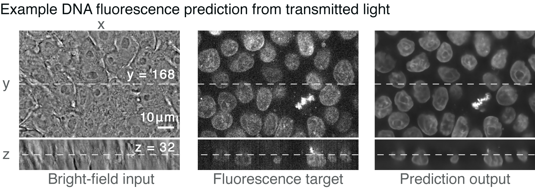
Fluorescence Microscopy Buyer's Guide - Biocompare Seeing is believing, and for many biologists, what they want to see is fluorescent labels through a microscope. Fluorescence microscopy, which makes it possible to visualize fluorescent proteins or dyes at the cellular and subcellular level, has become a workhorse of modern biology.. The concept of fluorescence, in which a molecule absorbs light of one wavelength and then emits light of a ... Click-ExM enables expansion microscopy for all biomolecules Dec 07, 2020 · Expansion microscopy (ExM) allows super-resolution imaging on conventional fluorescence microscopes, but has been limited to proteins and nucleic acids. ... Using 18 clickable labels, we ... Label-free prediction of three-dimensional fluorescence images from ... Label-free prediction of three-dimensional fluorescence images from transmitted-light microscopy Understanding cells as integrated systems is central to modern biology. Although fluorescence microscopy can resolve subcellular structure in living cells, it is expensive, is slow, and can damage cells.
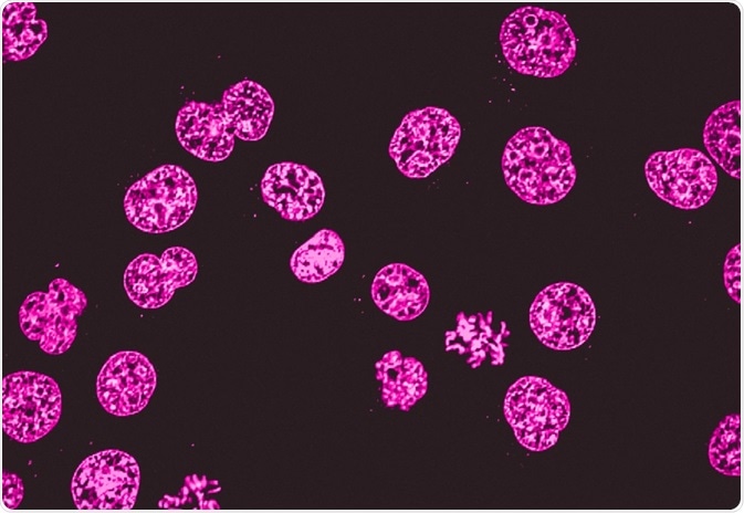
Fluorescent labels and light microscopy. Fluorescent Microscopy Fluorescent microscopy is often used to image specific features of small specimens such as microbes. It is also used to visually enhance 3-D features at small scales. This can be accomplished by attaching fluorescent tags to anti-bodies that in turn attach to targeted features, or by staining in a less specific manner. Fluorescence Live Cell Imaging - PMC - PubMed Central (PMC) Advantages are that full enclosures allow the best thermal equilibration. We always leave our system set to 37°C because it will take several hours for full temperature equilibration. This also reduces focus drift and minimizes thermal cycling experienced by optical and mechanical components. Fluorescent Label - an overview | ScienceDirect Topics 14.3.2 Fluorescence quenching microscopy Fluorescence microscopy is a very common tool. Usually, fluorescent labels are used to brighten up the object of interest. However, the same strategy is not applicable for graphitic materials, such as graphite, graphene, GO or r-GO as they are strong quenchers of dye molecules [45]. Fluorescent Dyes | Science Lab | Leica Microsystems In fluorescence microscopy there are two ways to visualize your protein of interest. Either with the help of an intrinsic fluorescent signal - by genetically linking a fluorescent protein to a target protein - or with the help of fluorescently labeled antibodies that bind specifically to a protein of interest.
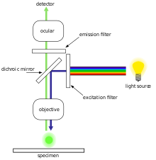
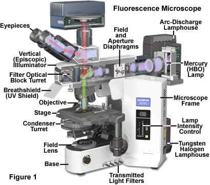
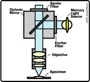
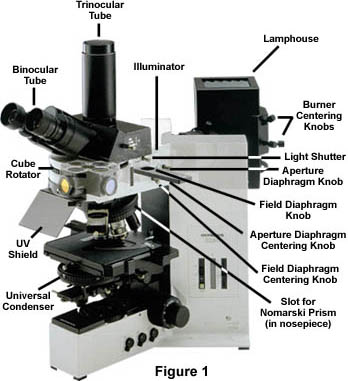
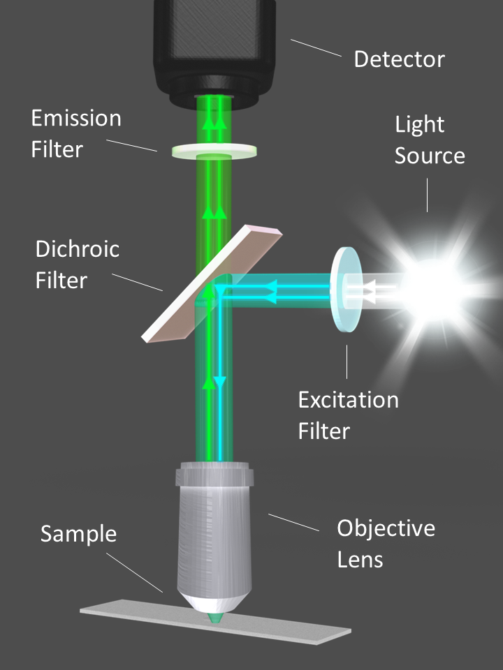
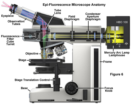
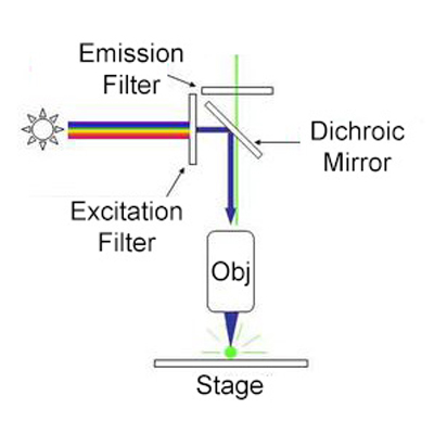
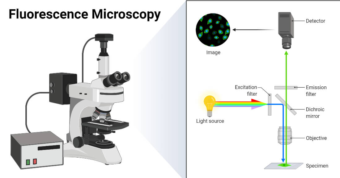
Förster resonance energy transfer - Wikipedia In fluorescence microscopy, fluorescence confocal laser scanning microscopy, as well as in molecular biology, FRET is a useful tool to quantify molecular dynamics in biophysics and biochemistry, such as protein-protein interactions, protein–DNA interactions, and protein conformational changes. For monitoring the complex formation between two ... Fluorescence microscope - Wikipedia Typical components of a fluorescence microscope are a light source ( xenon arc lamp or mercury-vapor lamp are common; more advanced forms are high-power LEDs and lasers ), the excitation filter, the dichroic mirror (or dichroic beamsplitter ), and the emission filter (see figure below). Anatomy of the Fluorescence Microscope - Olympus Reflected light fluorescence microscopes were first commercialized on a broad scale by Johan S. Ploem in the late 1960s, who was instrumental in developing the Wild-Leitz Ploem Opak, containing multiple optical blocks that were interchangeable and housed various combinations of filters for fluorescence microscopy. Fluorescent Labels - Neon Labels Inkjet/Laser | OnlineLabels® Our fluorescent label materials are bright, highly visible, neon colored labels with a permanent adhesive. The various five colors are ideal for signage, product labeling, organization, color coding and more. They have a matte finish that's great for printing logos, text, and designs and are both laser and inkjet printable for flexibility in ...
Geminate labels programmed by two-tone microdroplets ... - Nature Jan 29, 2021 · Guo and Li et al. recently have reported an interesting dual-mode CLC system induced by light-driven fluorescent chiral switches, in which the reflection wavelength and fluorescence intensity were ... Basics of FRET Microscopy | Nikon’s MicroscopyU The first fluorescent protein biosensor was a calcium indicator named cameleon, constructed by sandwiching the protein calmodulin and the calcium calmodulin-binding domain of myosin light chain kinase (M13 domain) between enhanced blue and green fluorescent proteins (EBFP and EGFP). In the presence of increasing levels of intracellular calcium ... EYFP :: Fluorescent Protein Database Note: There is inconsistency in the literature regarding the identity of "EYFP", and whether it contains V68L.Some papers (Lybarger et al. 1998; Patterson et al 2001) equate it with mutant 10C, introduced in Ormö et al 1996, which includes V68L. However, Miyawaki 1997 & 1999 and Nagai 2002 omit V68L, yet another paper from the same group (Rekas 2002) includes V68L (as … ProSciTech Laboratory supplies and Lab equipment for Histology, Pathology, Light Microscopy, Electron Microscopy and specialist researchers.
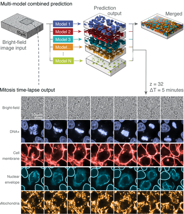
Label-free Determination - ALLEN CELL EXPLORER
Fluorescence Microscope: Principle, Types, Applications Fluorescence microscopy is a light microscope that works on the principle of fluorescence. A substance is said to be fluorescent when it absorbs the energy of invisible shorter wavelength radiation (such as UV light) and emits longer wavelength radiation of visible light (such as green or red light).

Fluorescence Microscopy - Cell Biology Flashcards | Draw it ...
Fluorescence - Wikipedia A perceptible example of fluorescence occurs when the absorbed radiation is in the ultraviolet region of the electromagnetic spectrum (invisible to the human eye), while the emitted light is in the visible region; this gives the fluorescent substance a distinct color that can only be seen when the substance has been exposed to UV light ...
ZEISS Microscopy Online Campus | Introduction to Spectral Imaging
Fluorescence or label-free imaging? Custom microscopy illumination ... For live cell imaging, label-free microscopy is a popular method which comes with several advantages. Unlike fluorescence microscopy, there are no labels that could potentially interfere with the phenomenon you want to observe. It also requires lower light levels, which is great for reducing photodamage.
Light microscopy of proteins in their ultrastructural context ...
Introduction to Fluorescence Microscopy | Nikon's MicroscopyU It is important to note that fluorescence is the only mode in optical microscopy where the specimen, subsequent to excitation, produces its own light. The emitted light re-radiates spherically in all directions, regardless of the excitation light source direction.

Optical Microscopy Application: Fluorescence | Edmund Optics
Fluorescence Microscopy vs. Light Microscopy - News-Medical.net This means that fluorescent microscopy uses reflected rather than transmitted light. For example, a commonly used label is green fluorescent protein (GFP), which is excited with blue...
Molecular Expressions Microscopy Primer: Specialized ...
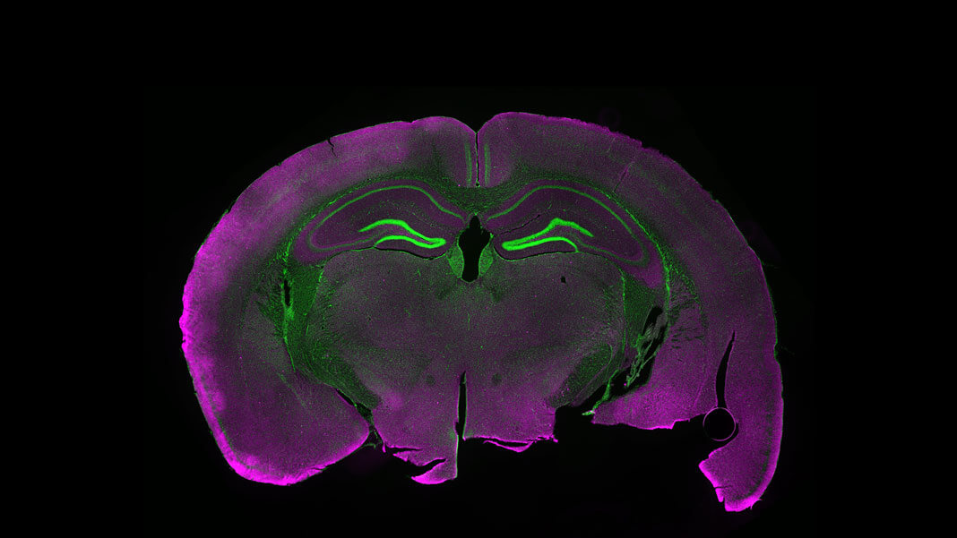
1.2 Fluorescence Light Microscopy - Cell Structure | Atlas The addition of fluorescence to light microscopy allows us to look not just at cells, but for things inside them. Specific cellular components can be fluorescently labeled, with a stain or antibody that binds a particular molecule.
Fluorescence Microscopy vs. Light Microscopy
Light Microscope- Definition, Principle, Types, Parts, Labeled Diagram ... A light microscope is a biology laboratory instrument or tool, that uses visible light to detect and magnify very small objects and enlarge them. They use lenses to focus light on the specimen, magnifying it thus producing an image. The specimen is normally placed close to the microscopic lens.

Feature-rich covalent stains for super-resolution and cleared ...
Fluorescence in Microscopy | Science Lab | Leica Microsystems Fluorescence in Microscopy. Fluorescence microscopy is a special form of light microscopy. It uses the ability of fluorochromes to emit light after being excited with light of a certain wavelength. Proteins of interest can be marked with such fluorochromes via antibody staining or tagging with fluorescent proteins.
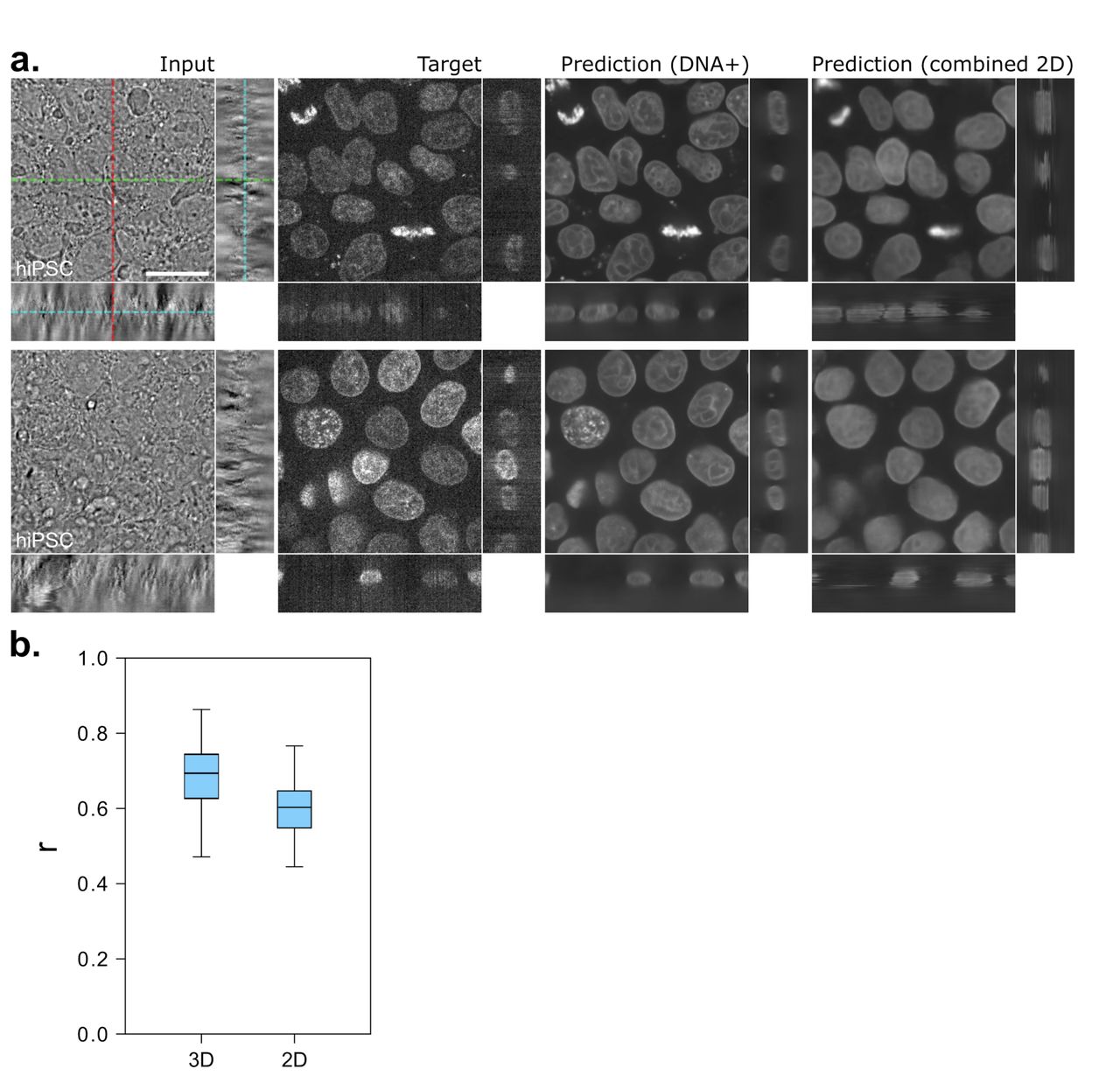
Label-free prediction of three-dimensional fluorescence ...
Fluorescent Labeling - What You Should Know - PromoCell Fluorescence microscopy allows the identification of cells and cellular components and the monitoring of cell physiology with high specificity. Fluorescence microscopy separates emitted light from excitation light using optical filters. The use of two indicators also allows the simultaneous observation of different biomolecules at the same time.
Fluorescence microscope - Wikipedia
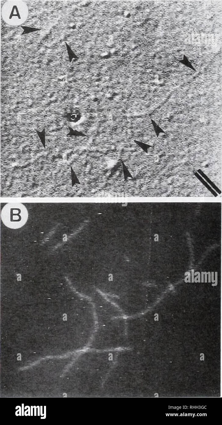
Fluorescent tag - Wikipedia S. cerevisiae septins revealed with fluorescent microscopy utilizing fluorescent labeling In molecular biology and biotechnology, a fluorescent tag, also known as a fluorescent label or fluorescent probe, is a molecule that is attached chemically to aid in the detection of a biomolecule such as a protein, antibody, or amino acid.
Fluorescence Microscopy - Anatomy of the Fluorescence ...
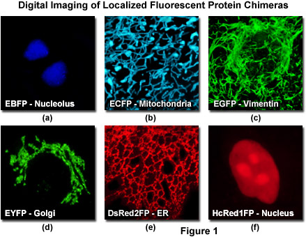
Dots, Probes and Proteins: Fluorescent Labels for Microscopy and Imaging GFP now comes in 'flavors' including cyan, yellow and blue. Fluorescent proteins are useful for studying live cells and can be used as 'reporters' for studying gene expression. Using genetically modified plasmid and/or viral DNA, the target cells can be transfected with the plasmid which encodes both the fluorescent protein and a gene ...
Hyperspectral multiphoton microscopy for in vivo ...
Why would scientists use fluorescent labels or dyes when using a light ... What is the advantage of fluorescence microscopy over light microscopy? Advantages of Fluorescence Microscope Fluorescence microscopy is the most popular method for studying the dynamic behavior exhibited in live-cell imaging. This stems from its ability to isolate individual proteins with a high degree of specificity amidst non-fluorescing ...

Fluorophores and Optical Filters for Fluorescence Microscopy
Fluorescence Microscopy - New York Microscope Company Fluorescence microscopy uses a high-intensity light source that excites a fluorescent molecule called a fluorophore in the sample observed. The samples are labeled with fluorophore where they absorb the high-intensity light from the source and emit a lower energy light of longer wavelength.
light-microscopy-fluorescent-labels-neuroscience-shutterstock ...
Label-free prediction of three-dimensional fluorescence images from ... We present a label-free method for predicting three-dimensional fluorescence directly from transmitted-light images and demonstrate that it can be used to generate multi-structure, integrated...
The Biological bulletin. Biology; Zoology; Biology; Marine ...
Light Sheet Fluorescence Microscopy | Nikon's MicroscopyU Light Sheet Fluorescence Microscopy ( LSFM) is a general name for a constantly growing family of planar illumination techniques that have revolutionized how optical imaging of biological specimens can be performed.
Fluorescence Microscopy - How Light Microscopes Work ...
Fluorescence Imaging - Teledyne Photometrics By targeting these fluorescent labels, researchers can select what they want to see. This is demonstrated in Fig.3, ... Two-photon Fluorescence Light Microscopy. Macmillan Publishing Group. Schermelleh, L., Heinztmann, R., and Leonardt, H. (2010). A Guide to Super-Resolution Fluorescence Microscopy. The Journal of Cell Biology 190 (2): 165-175.

The Microscope
Fluorescent labeling of abundant reactive entities (FLARE) for cleared ... Super-resolution microscopy Abstract Fluorescence microscopy is a vital tool in biomedical research but faces considerable challenges in achieving uniform or bright labeling. For instance,...

Simultaneous label-free autofluorescence-multiharmonic ...
Fluorescence Microscopy vs. Light Microscopy - New York Microscope Company Comparing Light vs Fluorescence Light microscopes use light in the 400-700nm range - the range through which light is visible to the human eye - but fluorescence microscopy uses much higher intensity light. Because traditional light microscopy uses visible light, the resolution is more limited.
Fluorescence - Reflected Light | Olympus LS
Different Ways to Add Fluorescent Labels - Thermo Fisher Scientific Labeling various targets with separate fluorescent colors allows you to visualize different structures or proteins within a cell in the same experiment. Ways to fluorescently label your target include fluorescent dyes, immunolabeling, and fluorescent fusion proteins —all of which can provide a means to selectively mark structures and proteins ...

Fluorescent tag - Wikipedia
Label-free prediction of three-dimensional fluorescence images from ... Label-free prediction of three-dimensional fluorescence images from transmitted-light microscopy Understanding cells as integrated systems is central to modern biology. Although fluorescence microscopy can resolve subcellular structure in living cells, it is expensive, is slow, and can damage cells.
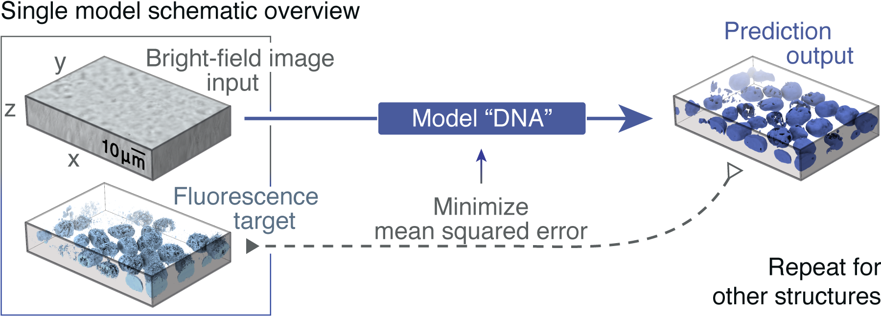
Label-free Determination - ALLEN CELL EXPLORER
Click-ExM enables expansion microscopy for all biomolecules Dec 07, 2020 · Expansion microscopy (ExM) allows super-resolution imaging on conventional fluorescence microscopes, but has been limited to proteins and nucleic acids. ... Using 18 clickable labels, we ...

Label-free imaging tool pipeline and application using 3D ...
Fluorescence Microscopy Buyer's Guide - Biocompare Seeing is believing, and for many biologists, what they want to see is fluorescent labels through a microscope. Fluorescence microscopy, which makes it possible to visualize fluorescent proteins or dyes at the cellular and subcellular level, has become a workhorse of modern biology.. The concept of fluorescence, in which a molecule absorbs light of one wavelength and then emits light of a ...
Labeling proteins inside living cells using external ...
Best practices and tools for reporting reproducible ...
Light Microscopy Techniques Used for Virology- Oxford Instruments

The Fundamentals and History of Fluorescence and Quantum Dots ...

Fluorescent Labeling - What You Should Know - PromoCell
Super-resolution fluorescence microscopy studies of human ...
Definition > Fluorescence microscope
Advanced Light Microscopy Methods - 2008 - Wiley Analytical ...
Fluorescence Filters for Microscopy and Imaging - Alluxa

Fluorescent microscopy - LNF Wiki

Practical Fluorescence Reconstruction Microscopy for High ...
Label-free Determination - ALLEN CELL EXPLORER

Fluorescent protein tags | Proteintech Group
ZEISS Microscopy Online Campus | Microscopy Basics ...
Fluorescence Microscopy - Explanation and Labelled Images ...
Fluorescence Microscopy- Definition, Principle, Parts, Uses

Fluorescent Microscopy

Bacterial Vivisection: How Fluorescence-Based Imaging ...

Cell Imaging
Hyperspectral multiphoton microscopy for in vivo ...
Fluorescence Microscopy Virtual Lab
Post a Comment for "43 fluorescent labels and light microscopy"